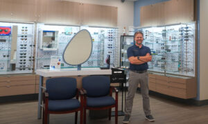
Multimodal diagnostic screening from Visionix, VX 120+ Dry Eye. Dr. Hoff says that next-generation innovation in eyecare is what enables practices to provide stellar care and differentiate themselves from competitors.
Sponsored Content
By Michelle J. Hoff, OD, FAAO, ABOM, FNAO
and Isabel Kazemi, OD, FAAO
August 2, 2023
In the past 20 years, technology has reshaped how we practice optometry.
Eyecare professionals who embrace technology and innovation increase access to eyecare and unlock the potential to elevate the patient experience.
Before we begin our journey to unlock your potential for increased patient care and profitability, we must define two concepts: disruptive innovation and technological advancement.
Disruptive innovation is making changes by integrating new technologies, streamlining processes and redefining how things are done, ultimately replacing existing modalities.
Technological advancement is a change in a product or service that is more precise and accurate, resulting in increased efficiency. Some technologies have been introduced into the eyecare space that have done this. Here are the details of how a suite of innovative instruments offered by Visionix can transform how practices can deliver an elevated patient care experience.
Why Do We Invest in New Technologies?
The digital phoropter is an appealing alternative to the manual phoropter, but hasn’t displaced the existing instrument or procedure. For better or worse, disruptive innovation has the potential to alter the standard of care, change the way we practice optometry, and will continue to influence our profession. In some cases, disruptive innovation represents a catalyst for positive change. Before adopting any new technology, we must evaluate how it applies to our primary goals.
Let’s take an example of a digital phoropter. Does this improve patient care and increase access to vision services? Will it help me focus more on the patient? Will this streamline patient flow and office efficiency? Is it easy to use? Will this investment boost practice revenue? What is the return on my investment?
An Integrated Technology Suite with Visual Tools that’s a Game-Changer
Visionix technologies provide precisely what we are talking about regarding positive technological innovation and disruption.
Let’s present the instruments in the order of the exam flow.
The first piece of data to collect is usually the spectacle Rx.
Compared to a manual or automated lensometer, the VX 40 lens analyzer is fully automated, measures a larger lens area, and uses wavefront technology to create three topographic maps.
Unlike the manual and automated lensometers, which are user dependent and require a skilled technician to obtain reliable measurements, the VX 40 eliminates user dependence and captures reliable, repeatable data. The instrument automatically identifies the difference between single-vision and variable-power lenses.
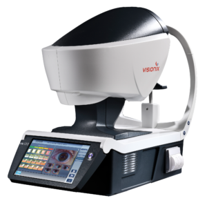
Next, the technician would use the fully automated VX 120+ Dry Eye or VX 650 diagnostic unit to complete a visual analysis in around two minutes. These multimodal devices perform auto refraction, keratometry, aberrometry, topography, pachymetry, tonometry, angle evaluation and more.
The main difference between these two instruments is that the VX 120+DE has the dry eye module, and the VX 650 has the retinal imaging module, allowing you to select the unit that best meets your practice needs.
As we move through the exam flow, remember that the pre-examination equipment has already provided you with comprehensive diagnostic data.
The VX 120+ DE is an ideal diagnostic tool for dry eye specialty practices and collects medically billable dry eye diagnostic data. It features analysis of tear break-up time, tear meniscus height and high-definition color imaging of the meibomian glands.
With a Dry Eye module and automated tonometry, there is no need for fluorescein, which saves you time and is a better experience for your patient.
The VX 650, with the retinal imaging module, is an ideal diagnostic tool for posterior segment specialty practices and collects medically billable posterior segment diagnostic data to detect early signs of glaucoma, diabetic changes, AMD and other retinal pathologies.
Using all the diagnostic tools from these instruments, you can review the images with your patients to demonstrate the need for a personalized treatment plan and track their progress.
The Eye Refract is a unique instrument that can be used in the pre-exam or exam room.
In the pre-examination area, the technician conducts a majority of the visual assessment, including data collection from the instrument’s subjective refinement, then transmits the findings to the exam room for the doctor to further refine (if needed), which significantly reduces the patient’s time behind the phoropter.
The patient’s vision is measured under bright and low light conditions to generate day and nighttime prescriptions, and this is unique to the Eye Refract. Measurements are taken under binocular conditions using two wavefront aberrometers and a fully automated digital phoropter to deliver a highly accurate visual assessment in an average of four minutes.
The traditional refraction process, which includes objective, subjective measurements, a binocular balance and near testing, takes about 10 minutes.
Improve Patient Care & Boost Profitability
Investment in technology is driven by a desire to improve efficiency, provide the highest quality of patient care and slow disease progress. The investment touches all the pain points of the practice flow, collecting valuable diagnostic data throughout the process.
New automated technologies facilitate information gathering and documenting at the highest levels, streamlining office efficiency and transforming each step of your process. Each point of contact during the patient’s visit is an opportunity to build trust and gain patient loyalty.
For example, the iWellness Exam is a unique technology in all Optovue OCTs by Visionix, which provides early detection of many ocular diseases and flags existing pathology 96 percent of the time.
If you offer screening fundus photos, you are already familiar with a wellness screening; however, the OCT is an advanced three-dimensional, high-resolution approach, elevating patient care to a higher standard.
It can be a new revenue source because it is a separate charge from the medically billable OCT imaging.
For medically billable OCT imaging, a variety of advanced instruments are available to practices that provide specialty care, such as dry eye, glaucoma or retinal disease management. These multi-diagnostic instruments offer state-of-the-art imaging from cornea to choroid.
Furthermore, adding or updating existing lab finishing equipment to state-of-the-art machines will reduce your lab bill, increase profits and differentiate you from other practices.
The Briot Couture Lens Finishing system by Visionix includes an integrated space-saving blocker and edger. The blocking unit uses wavefront technology to preview the lens design, automatically and accurately blocking the lens, eliminating the need for lensometry skills. This is the only tracer on the market able to create a three-dimensional model of the entire frame, including thickness, shape and groove placement. Three-dimensional graphic visualization lets the optician and the patient preview the predicted finished eyewear.
Other Articles to Explore
This feature is extremely useful for the optician when selecting the most appropriate frame, lens material and lens design, especially when designing eyewear for challenging prescriptions.
The advanced innovations in the machine’s sensors and software design eliminate the biggest challenges in edging to reduce lens remakes significantly.
These edgers are environmentally friendly because the water consumption has been considerably reduced from 16 liters to five. An in-office finishing lab elevates the patient’s vision care level because you can offer faster delivery or same-day prescriptions, adding a new source of revenue to the practice.
Elevate the Patient Experience, Keep Up with Modern Expectations
Today’s patients are more proactive and involved in their healthcare and expect more from their providers.
Whether the problem is due to ophthalmic lenses or anterior or posterior ocular conditions, sharing visual data with the patient involves them in their own vision care and, thus, motivates better compliance with your treatment plan.
Investing in a comprehensive portfolio of technologically advanced instruments, like that provided by Visionix, improves your ability to communicate complex findings and elevates the quality of care received in your office. Through the experience of an impressive level of technological innovation in-house, patients will know you’re the first place they should turn to for “all things eye.”
 Michelle J. Hoff, OD, FAAO, ABOM, FNAO, is an Associate Clinical Professor at Herbert Wertheim School of Optometry & Vision Science, University of California Berkeley.
 Isabel Kazemi, OD, FAAO, is an Assistant Clinical Professor at Herbert Wertheim School of Optometry & Vision Science, University of California Berkeley.