Technology to transform the care of your patients with diabetes.
Sponsored Content
By Bobby W. Wood, OD
Nov. 1, 2023
If you came to me two years ago and told me that a handheld ERG device was going to help me better manage my patients with diabetes and boost my bottom line, I’m not sure I would have believed you, but I experienced it first-hand.
The RETeval ERG from LKC Technologies is a game-changing handheld full-field ERG device that allows optometrists to gather functional data to better manage their patients with diabetes at a fraction of the cost of a spectral domain OCT or wide-field fundus camera—and with fair reimbursements. When I first started learning about this, I thought it sounded too good to be true, but now that I’ve integrated this technology into my practice—quite seamlessly—I firmly believe that the RETeval full-field ERG test for functional diabetic retinopathy management will change the way we practice in 2024 in much the same way that OCT testing for structural diabetic retinopathy management changed the way we practiced 20 years ago.
How Does ERG Work?
Electroretinography (ERG) measures the electrical responses of various cell types in the retina, including the photoreceptors (rods and cones), inner retinal cells (bipolar and amacrine cells) and the ganglion cells in response to a stimulus. These stimuli can be measured as a waveform stimulus as light passes through the eye and onto the retina. The retinal photoreceptors (rods and cones) absorb the light before converting the stimulus. This is known as the A-wave or Implicit Time. A delay in Implicit Time (abnormal A wave) indicates cellular stress.
Next, the light gets converted into electrochemical impulses that get transmitted to the bipolar cells. This is known as the B-wave or Amplitude. A reduced Amplitude (abnormal B Wave) indicates cells are dying or the number of healthy cells is decreasing. The bipolar cells effectively transfer information from rods and cones to the retinal ganglion cells, which are responsible for carrying visual information from the eye to the brain, this is known as the PhNR wave. An abnormal PhNR is indicative of reduced function of the retinal ganglion cells and is most common in conditions that affect the optic nerve, including ischemic optic neuropathy and glaucoma.
Is ERG Practical?
Historically, due to the size and complexity of ERG equipment and the challenge of interpreting the data, ERG was reserved for research centers, hospitals and academic facilities, but now thanks to the handheld RETeval device, ERG testing is available for all eyecare providers. The RETeval simplifies the application and interpretation of ERG by providing:
- Handheld, mobile unit that can be used anywhere in the office
- Constant pupil tracking that allows for non-dilated testing
- Patient-friendly
- Staff-friendly administration
- An easy user interface in multiple languages
- Color-coded result interpretation
- A “Diabetic Retinopathy Score”
- Testing that can be completed in minutes
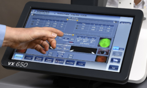
ERG testing with the RETeval is simple and only takes a few minutes thanks to FDA-cleared adhesive skin electrodes that are placed under each eye providing improved patient comfort since no corneal contact is required. The sensor strips connect to the handheld device with a quick and easy wire clip attachment. Each sensor strip contains three electrodes to reduce placement variability.
Constant pupil tracking allows for reliable, repeatable results with or without dilation. The device dynamically changes the stimulus intensity according to the pupil size to deliver a constant retinal illuminance during testing, which reduces the variability of measurement results caused by pupil size.
The “secret sauce” that really excited me clinically about the RETeval ERG is that it is an OBJECTIVE TEST of RETINAL FUNCTION that is PREDICTIVE in diagnosing retinal eye disease. It provides objective functional test results that are successful and reliable, even in challenging cases, such as in patients who are uncooperative (i.e., small children, TBI, dementia) or who have media opacities, including cataracts.
My Clinical Experience with ERG
So how does RETeval ERG help me better manage my patients with diabetic retinopathy? My favorite feature of the device is the DR Assessment Protocol, which produces a DR Score (diabetic retinopathy score), and can help predict whether a patient may require diabetic retinopathy intervention treatment. This score is determined by four parameters: implicit time, amplitude, pupil response and patient age. We know that ischemic changes of the iris can cause reduced or sluggish pupil responses over time in patients with diabetes. By taking the best eye implicit time and amplitude and combining it with the worse eye pupil response plus patient age, a DR Score is determined.
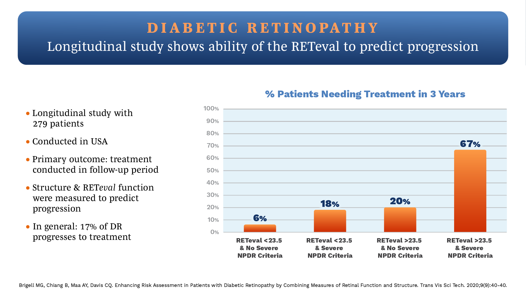
As seen in the graph above, if a patient’s RETeval DR Score is less than 23.5, the risk of surgical intervention in the next three years varies from 6 percent to 18 percent, depending upon their level of non-proliferative diabetic retinopathy (NPDR). If the patient’s DR Score is greater than 23.5 and they have severe NPDR, the risk of surgical intervention in the next three years increases to 67 percent. With a DR Score over 26, surgical intervention may be needed within one year.
The DR Score can worsen with inadequate diabetic control, but it can also improve with successful diabetic management. By educating the patient and their diabetic healthcare provider of their diabetic retinopathy status and RETeval DR Score, we can assist the diabetic healthcare provider in managing our patient’s diabetes and reduce the risk for referral to a retinal specialist for surgical intervention and potential vision loss.
Read Another Success Story
Thanks to functional testing with the RETeval ERG, when combined with structural testing with dilated retinal examination, UWF fundus photography and SD-OCT, I am now able to DETECT retinal disease earlier, PREDICT the progression of the disease, FOLLOW-up on the course of the disease and MONITOR treatment success for my patients with diabetes in a way that was not possible in the past, but now is thanks to the RETeval ERG with DR Score.
Is ERG Profitable?
The RETeval helps me address the retinal health of my patients, but it also boosts the bottom line and financial health of our practices in the following ways:
- Reimbursement: The local Medicare reimbursement in San Antonio where I practice for code 92273 (full field ERG) is $122.66 (the national average reimbursement of $128.09 as a bilateral test). Compare that to 92250 (fundus photography) at $36.39 and 92134 (OCT of the retina) at $39.77. Best of all, there are no competing codes, so fundus photography, OCT and/or visual fields can be performed and reimbursed on the same day as RETeval ERG with one of more than 560 approved ICD-10 codes.
- Equipment cost: The RETeval ERG is very cost effective compared to other diagnostic equipment. The cost of a RETeval device is in the low-to-mid 20s. Compare that to the cost of a SD-OCT or UWF fundus camera. Lower entry costs help with cash flow.
- ROI: Thanks to the reimbursement rate and low relative equipment cost of the RETeval ERG, the return on investment (ROI) is extremely fast. If a practice performs two RETeval ERG tests per day, the practice will earn back their ROI in approximately four months.
I have two practices with a RETeval ERG device at each location. We use them every day, primarily for our patients with diabetic retinopathy. With the small footprint of the device, the smooth integration of implementing this test into our daily clinic schedule, the ease of use by our technicians and the easy-to-read and interpret color-coded exam reports for our doctors, the RETeval ERG has most definitely been a fantastic addition for diabetes management in our clinic and is definitely not only good, but GREAT for both patient care and for the practice bottom line.
 Bobby W. Wood, OD, is the owner of Wood Vision Source in San Antonio, Texas.