Sept. 20, 2017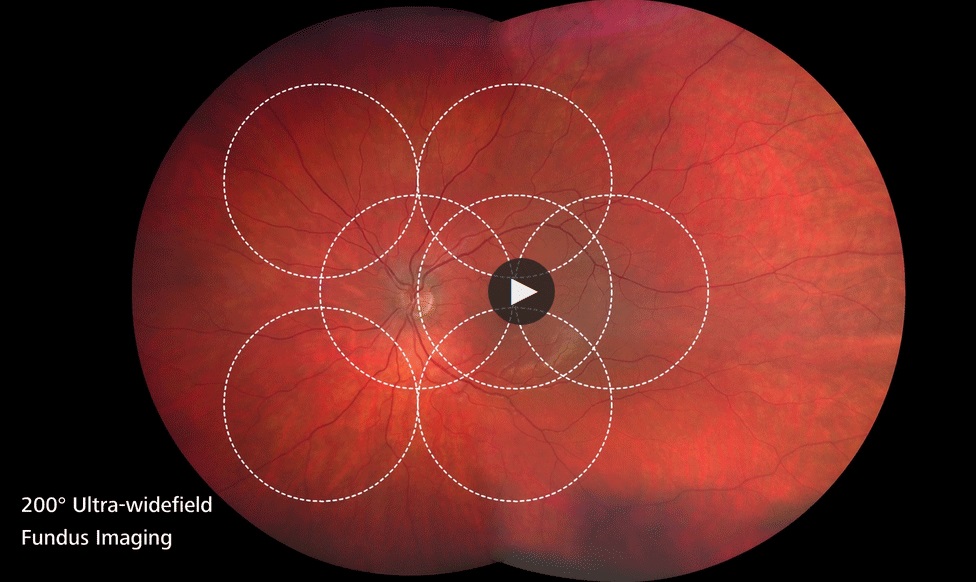
ZEISS Medical Technology announced the U.S. launch of its next generation, high-definition Ultra-widefield (HD UWF) fundus imaging system, CLARUS 500, at the International Vision Expo & Conference in Las Vegas, Nev.
Leveraging ZEISS precision optics, ZEISS says the CLARUS 500 is the first fundus camera that combines true color with high-resolution clarity down to 7 microns in an Ultra-widefield view from the macula to the far periphery. Early signs of eye disease can often be subtle, and can occur in the far periphery of the retina.
This new ZEISS system is designed to allow doctors to obtain a better view of the entire retina. CLARUS 500 produces images in True Color that closely resemble the coloration of the retina as seen through direct observation during clinical examination.
Color accuracy is important in the diagnosis, documentation and management of ocular diseases; ensuring confidence when evaluating optic disc, nevi, and lesions in which subtle color differences may lead to a change in diagnosis and management. Specifically, accurate coloration and resolution is important for evaluating focal change in rim tissue, nerve pallor, dry age-related macular degeneration RPE pigment changes, and drusen.
According to retinal specialist, Roger Goldberg MD, MBA with Bay Area Retina Associates, CLARUS 500 from ZEISS combines the benefits of an ultra-widefield view across the entire retina with the high-quality of traditional fundus photography used for imaging the optic nerve and macula. “By offering true-color, high-resolution images of the optic nerve and macula, as well as the retinal periphery, we no longer have to choose which system to image a patient on based on their retinal pathology,” Dr. Goldberg added.
“Traditional fundus imaging systems have been the gold standard for macular disease diagnosis and optic nerve evaluation for many years,” says Jim Mazzo, Global President Ophthalmic Devices at Carl Zeiss Meditec. “Now, Ultra-widefield is starting to change this. Clinicians are finding that by imaging a larger area of the retina, they have the possibility of uncovering more pathology, aiding in earlier disease diagnosis and better patient management. With ZEISS CLARUS, they can better manage a broader range of patients with one fundus imaging system,” Mazzo continues.
With a single capture, ZEISS CLARUS 500 produces a 133-degree HD widefield image. HD widefield images are automatically merged to achieve a 200-degree ultra-widefield of view. The technology allows clinicians to easily review and compare high-quality images captured during a single exam while providing annotation and caliper measurement tools that allow in-depth analysis of eye health. CLARUS integrates with ZEISS FORUM and Retina Workplace for review with other ophthalmic images and exam data for efficient multi-modality analysis.
Another advantage of the CLARUS 500 is its ability for peripheral imaging while still maintaining the ability to zoom into the retina without losing resolution. Reticula degeneration in the periphery is a risk factor for AMD progression.
“ZEISS continually strives to evolve our legendary precision optics technology to help doctors advance patient care,” said Dr. Ludwin Monz, President and CEO of Carl Zeiss Meditec. “We are pleased to be able to add advanced Ultra-widefield imaging to the comprehensive portfolio of diagnostic imaging solutions that help doctors more easily detect eye disease and help preserve their patient’s vision.”
ZEISS CLARUS 500 Ultra-widefield is available in the United States beginning in September 2017. Availability is pending in all other major markets.
For more information about ZEISS CLARUS 500, visit www.zeiss.com/us/clarus